Figure 7
Stroke Mimics: Insights from a Retrospective Neuroimaging Study
Lucia Monti*, Davide del Roscio, Francesca Tutino, Tommaso Casseri, Umberto Arrigucci, Matteo Bellini, Maurizio Acampa, Sabina Bartalini, Carla Battisti, Giovanni Bova and Alessandro Rossi
Published: 27 September, 2023 | Volume 7 - Issue 2 | Pages: 094-103
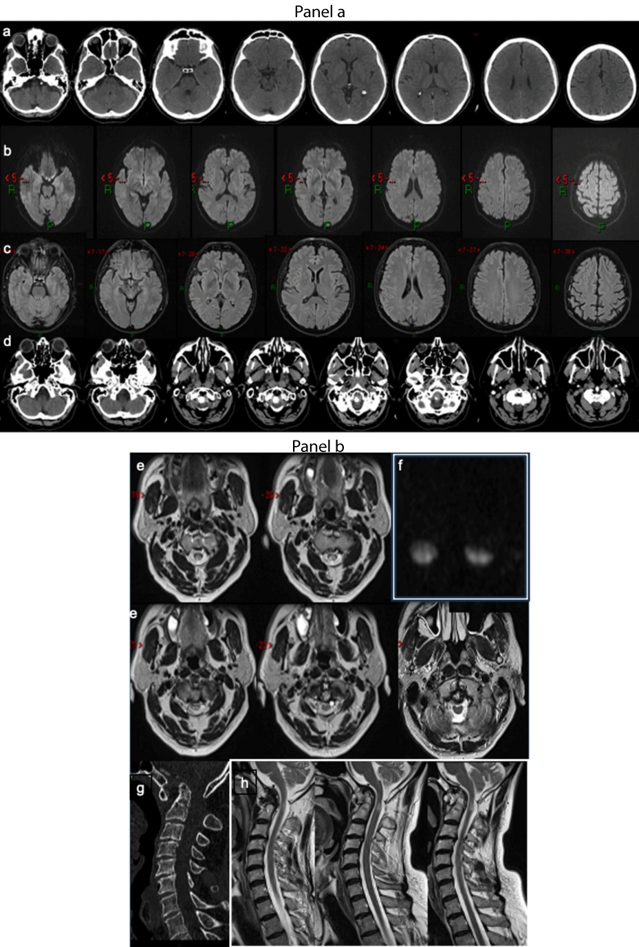
Figure 7:
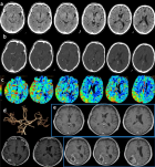
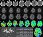
Figure 7: Spinal cord compression. Panel a. Stroke code: 58 years old male presenting with suddenly augmentation right upper limb paresis and paraesthesia. (a) No-contrast brain CT and intracranial CTA (not showed) did not reveal any vascular or parenchymal abnormalities or subacute ischemic lesions MRI did not demonstrate brain areas of restricted diffusion were evident on (b) DWI or FLAIR (c) signal alteration. The non-contrast CT of the cranio-cervical tract shows the odontoid process dislocated superiorly with involvement of the foramen magnum.(d) Panel b. Axial T2-weighted MRI images show “owl-eyes” sign at C1/C2 level (e). Intramedullary lesions at C1/C2 level were confirmed diffusion images (f).Cervical spine CT and sagittal reconstruction (g) and T2 sagittal images revealed an odontoid process subluxation and the focal hyperintensity of cervical spinal cord (h) Diagnosis: Cervical myelopathy. Atlantoaxial instability for degenerative changes caused the spinal cord compression and acute myelopathy.
Read Full Article HTML DOI: 10.29328/journal.jnnd.1001083 Cite this Article Read Full Article PDF
More Images
Similar Articles
-
Analysis of early Versus Delayed Carotid Surgery after Acute Ischemic StrokePEROU Sébastien*,DETANTE Olivier,SPEAR Rafaelle,PIRVU Augustin,ELIE Amandine,MAGNE Jean-Luc. Analysis of early Versus Delayed Carotid Surgery after Acute Ischemic Stroke. . 2017 doi: 10.29328/journal.jnnd.1001001; 1: 001-011
-
Endovascular treatment experience in acute ischemic strokeİbrahim Acır*,Vildan Yayla,Hacı Ali Erdoğan. Endovascular treatment experience in acute ischemic stroke. . 2021 doi: 10.29328/journal.jnnd.1001047; 5: 026-028
-
Endovascular management of tandem occlusions in stroke: Treatment strategies in a real-world scenarioJuan J Cirio*,Celina Ciardi,Matias Lopez,Esteban V Scrivano,Javier Lundquist,Ivan Lylyk,Nicolas Perez,Pedro Lylyk. Endovascular management of tandem occlusions in stroke: Treatment strategies in a real-world scenario. . 2021 doi: 10.29328/journal.jnnd.1001051; 5: 055-060
-
Stroke Mimics: Insights from a Retrospective Neuroimaging StudyLucia Monti*, Davide del Roscio, Francesca Tutino, Tommaso Casseri, Umberto Arrigucci, Matteo Bellini, Maurizio Acampa, Sabina Bartalini, Carla Battisti, Giovanni Bova, Alessandro Rossi. Stroke Mimics: Insights from a Retrospective Neuroimaging Study. . 2023 doi: 10.29328/journal.jnnd.1001083; 7: 094-103
Recently Viewed
-
Chronic endometritis in in vitro fertilization failure patientsAfaf T Elnashar*,Mohamed Sabry. Chronic endometritis in in vitro fertilization failure patients. Clin J Obstet Gynecol. 2020: doi: 10.29328/journal.cjog.1001073; 3: 175-181
-
Relation of Arachnophobia with ABO blood group systemMuhammad Imran Qadir,Sani E Zahra*. Relation of Arachnophobia with ABO blood group system. J Hematol Clin Res. 2019: doi: 10.29328/journal.jhcr.1001011; 3: 050-052
-
Preservation of Haemostasis with Anti-thrombotic Serotonin AntagonismMark IM Noble*,Angela J Drake-Holland. Preservation of Haemostasis with Anti-thrombotic Serotonin Antagonism. J Hematol Clin Res. 2017: doi: 10.29328/journal.jhcr.1001004; 1: 019-025
-
Neutrophil to Lymphocyte Ratio (NLR) in Peripheral Blood: A Novel and Simple Prognostic Predictor of Non-small Cell Lung Cancer (NSCLC)Xiaoli Zhang,Ziyuan Zou,Liyu Fan,Xinjie Xu,Yu Siyuan,Peng Luo*. Neutrophil to Lymphocyte Ratio (NLR) in Peripheral Blood: A Novel and Simple Prognostic Predictor of Non-small Cell Lung Cancer (NSCLC). J Hematol Clin Res. 2017: doi: 10.29328/journal.jhcr.1001002; 1: 011-013
-
Gilbert’s Syndrome Revealed by Hepatotoxicity of ImatinibImen Ben Amor*,Imen Frikha,Moez Medhaffer,Moez Elloumi. Gilbert’s Syndrome Revealed by Hepatotoxicity of Imatinib. Ann Clin Gastroenterol Hepatol. 2025: doi: 10.29328/journal.acgh.1001049; 9: 001-003
Most Viewed
-
Evaluation of Biostimulants Based on Recovered Protein Hydrolysates from Animal By-products as Plant Growth EnhancersH Pérez-Aguilar*, M Lacruz-Asaro, F Arán-Ais. Evaluation of Biostimulants Based on Recovered Protein Hydrolysates from Animal By-products as Plant Growth Enhancers. J Plant Sci Phytopathol. 2023 doi: 10.29328/journal.jpsp.1001104; 7: 042-047
-
Sinonasal Myxoma Extending into the Orbit in a 4-Year Old: A Case PresentationJulian A Purrinos*, Ramzi Younis. Sinonasal Myxoma Extending into the Orbit in a 4-Year Old: A Case Presentation. Arch Case Rep. 2024 doi: 10.29328/journal.acr.1001099; 8: 075-077
-
Feasibility study of magnetic sensing for detecting single-neuron action potentialsDenis Tonini,Kai Wu,Renata Saha,Jian-Ping Wang*. Feasibility study of magnetic sensing for detecting single-neuron action potentials. Ann Biomed Sci Eng. 2022 doi: 10.29328/journal.abse.1001018; 6: 019-029
-
Pediatric Dysgerminoma: Unveiling a Rare Ovarian TumorFaten Limaiem*, Khalil Saffar, Ahmed Halouani. Pediatric Dysgerminoma: Unveiling a Rare Ovarian Tumor. Arch Case Rep. 2024 doi: 10.29328/journal.acr.1001087; 8: 010-013
-
Physical activity can change the physiological and psychological circumstances during COVID-19 pandemic: A narrative reviewKhashayar Maroufi*. Physical activity can change the physiological and psychological circumstances during COVID-19 pandemic: A narrative review. J Sports Med Ther. 2021 doi: 10.29328/journal.jsmt.1001051; 6: 001-007

HSPI: We're glad you're here. Please click "create a new Query" if you are a new visitor to our website and need further information from us.
If you are already a member of our network and need to keep track of any developments regarding a question you have already submitted, click "take me to my Query."


