Figure 4
Stroke Mimics: Insights from a Retrospective Neuroimaging Study
Lucia Monti*, Davide del Roscio, Francesca Tutino, Tommaso Casseri, Umberto Arrigucci, Matteo Bellini, Maurizio Acampa, Sabina Bartalini, Carla Battisti, Giovanni Bova and Alessandro Rossi
Published: 27 September, 2023 | Volume 7 - Issue 2 | Pages: 094-103
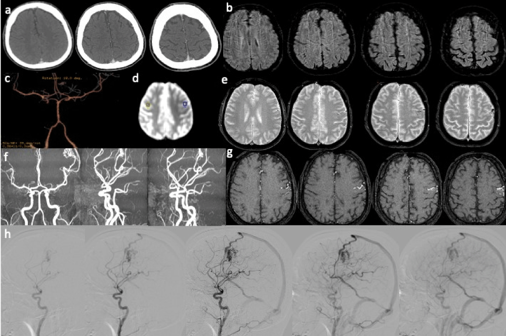
Figure 4:
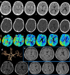
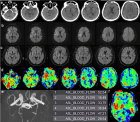
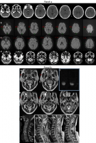
Figure 4: Arteriovenous malformation. Acute focal neurologic symptoms. Stroke code: 45 years old male presenting with right hemiparesis, weakness, impairment of speech and alteration of consciousness (NIHSS 6). a) No-contrast brain CT and b) CTA findings were negative. d) T2- weighted MRI shows ectasic surface vessels in the left frontal lobe, better appreciated on (e) SWI. f) Quantitative ASL (OLEA sphere 3.0): focal hypoperfusion corresponding to the left Rolandic cortical area adjacent to ectasic cortical vessels. 3D-TOF MRA(g,h) (single slice and MIP) shows the abnormal dilated vessels embedded left frontal lobe and (f) the DSA on lateral left carotid angiograms demonstrate the left frontal AVM with a nidus supplied by an enlarged branch of MCA. Note the early drainage to a frontal cortical vein into the superior sagittal sinus (and via an internal cerebral vein to the straight sinus). Diagnosis: frontal AVM inducing focal hypoperfusion of left Rolandic cortical area caused the acute neurologic symptoms.
Read Full Article HTML DOI: 10.29328/journal.jnnd.1001083 Cite this Article Read Full Article PDF
More Images
Similar Articles
-
Analysis of early Versus Delayed Carotid Surgery after Acute Ischemic StrokePEROU Sébastien*,DETANTE Olivier,SPEAR Rafaelle,PIRVU Augustin,ELIE Amandine,MAGNE Jean-Luc. Analysis of early Versus Delayed Carotid Surgery after Acute Ischemic Stroke. . 2017 doi: 10.29328/journal.jnnd.1001001; 1: 001-011
-
Endovascular treatment experience in acute ischemic strokeİbrahim Acır*,Vildan Yayla,Hacı Ali Erdoğan. Endovascular treatment experience in acute ischemic stroke. . 2021 doi: 10.29328/journal.jnnd.1001047; 5: 026-028
-
Endovascular management of tandem occlusions in stroke: Treatment strategies in a real-world scenarioJuan J Cirio*,Celina Ciardi,Matias Lopez,Esteban V Scrivano,Javier Lundquist,Ivan Lylyk,Nicolas Perez,Pedro Lylyk. Endovascular management of tandem occlusions in stroke: Treatment strategies in a real-world scenario. . 2021 doi: 10.29328/journal.jnnd.1001051; 5: 055-060
-
Stroke Mimics: Insights from a Retrospective Neuroimaging StudyLucia Monti*, Davide del Roscio, Francesca Tutino, Tommaso Casseri, Umberto Arrigucci, Matteo Bellini, Maurizio Acampa, Sabina Bartalini, Carla Battisti, Giovanni Bova, Alessandro Rossi. Stroke Mimics: Insights from a Retrospective Neuroimaging Study. . 2023 doi: 10.29328/journal.jnnd.1001083; 7: 094-103
Recently Viewed
-
Chronic endometritis in in vitro fertilization failure patientsAfaf T Elnashar*,Mohamed Sabry. Chronic endometritis in in vitro fertilization failure patients. Clin J Obstet Gynecol. 2020: doi: 10.29328/journal.cjog.1001073; 3: 175-181
-
Relation of Arachnophobia with ABO blood group systemMuhammad Imran Qadir,Sani E Zahra*. Relation of Arachnophobia with ABO blood group system. J Hematol Clin Res. 2019: doi: 10.29328/journal.jhcr.1001011; 3: 050-052
-
Preservation of Haemostasis with Anti-thrombotic Serotonin AntagonismMark IM Noble*,Angela J Drake-Holland. Preservation of Haemostasis with Anti-thrombotic Serotonin Antagonism. J Hematol Clin Res. 2017: doi: 10.29328/journal.jhcr.1001004; 1: 019-025
-
Neutrophil to Lymphocyte Ratio (NLR) in Peripheral Blood: A Novel and Simple Prognostic Predictor of Non-small Cell Lung Cancer (NSCLC)Xiaoli Zhang,Ziyuan Zou,Liyu Fan,Xinjie Xu,Yu Siyuan,Peng Luo*. Neutrophil to Lymphocyte Ratio (NLR) in Peripheral Blood: A Novel and Simple Prognostic Predictor of Non-small Cell Lung Cancer (NSCLC). J Hematol Clin Res. 2017: doi: 10.29328/journal.jhcr.1001002; 1: 011-013
-
Gilbert’s Syndrome Revealed by Hepatotoxicity of ImatinibImen Ben Amor*,Imen Frikha,Moez Medhaffer,Moez Elloumi. Gilbert’s Syndrome Revealed by Hepatotoxicity of Imatinib. Ann Clin Gastroenterol Hepatol. 2025: doi: 10.29328/journal.acgh.1001049; 9: 001-003
Most Viewed
-
Evaluation of Biostimulants Based on Recovered Protein Hydrolysates from Animal By-products as Plant Growth EnhancersH Pérez-Aguilar*, M Lacruz-Asaro, F Arán-Ais. Evaluation of Biostimulants Based on Recovered Protein Hydrolysates from Animal By-products as Plant Growth Enhancers. J Plant Sci Phytopathol. 2023 doi: 10.29328/journal.jpsp.1001104; 7: 042-047
-
Sinonasal Myxoma Extending into the Orbit in a 4-Year Old: A Case PresentationJulian A Purrinos*, Ramzi Younis. Sinonasal Myxoma Extending into the Orbit in a 4-Year Old: A Case Presentation. Arch Case Rep. 2024 doi: 10.29328/journal.acr.1001099; 8: 075-077
-
Feasibility study of magnetic sensing for detecting single-neuron action potentialsDenis Tonini,Kai Wu,Renata Saha,Jian-Ping Wang*. Feasibility study of magnetic sensing for detecting single-neuron action potentials. Ann Biomed Sci Eng. 2022 doi: 10.29328/journal.abse.1001018; 6: 019-029
-
Pediatric Dysgerminoma: Unveiling a Rare Ovarian TumorFaten Limaiem*, Khalil Saffar, Ahmed Halouani. Pediatric Dysgerminoma: Unveiling a Rare Ovarian Tumor. Arch Case Rep. 2024 doi: 10.29328/journal.acr.1001087; 8: 010-013
-
Physical activity can change the physiological and psychological circumstances during COVID-19 pandemic: A narrative reviewKhashayar Maroufi*. Physical activity can change the physiological and psychological circumstances during COVID-19 pandemic: A narrative review. J Sports Med Ther. 2021 doi: 10.29328/journal.jsmt.1001051; 6: 001-007

HSPI: We're glad you're here. Please click "create a new Query" if you are a new visitor to our website and need further information from us.
If you are already a member of our network and need to keep track of any developments regarding a question you have already submitted, click "take me to my Query."


