Figure 2
Intrasellar psammomatous meningioma: a case report and review of the literature
Luca Riccioni*, Antonio Balestrieri, Fuschillo Dalila, Maria Teresa Nasi and Luigino Tosatto
Published: 18 January, 2022 | Volume 6 - Issue 1 | Pages: 011-015

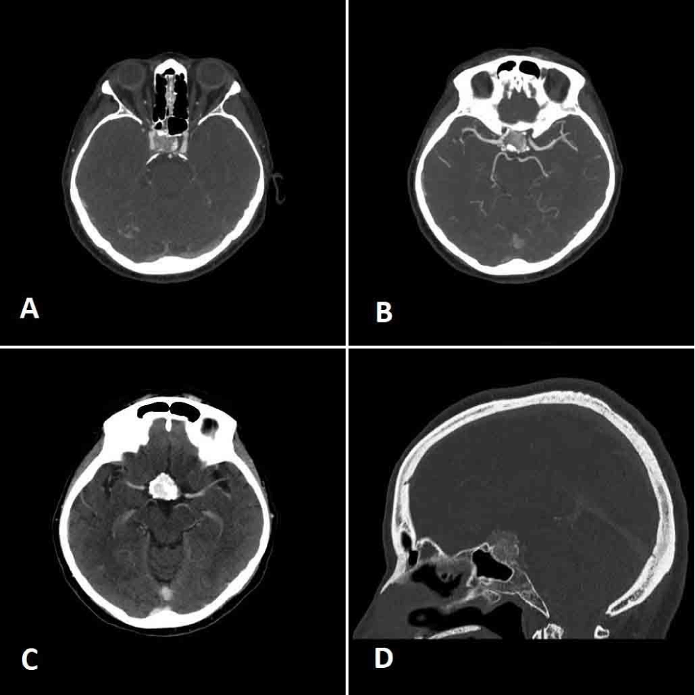
Figure 2:
A. and B. Axial angio-CT scan demonstrating the encasement of the right carotid artery, the presence of calcifications and the moderate sellar enlargement; C. axial CT scan showing the early and marked contrast enhancement of the mass; D. sagittal CT scan showing the thinning of the sellar floor due to the expansive mass.
Read Full Article HTML DOI: 10.29328/journal.jnnd.1001061 Cite this Article Read Full Article PDF
More Images
Similar Articles
-
Pituitary adenoma and meningioma simulating a single selar and paraseal injuryCarlos Pérez-López*,Alexis J Palpán Flores,Sharona Azriel,Víctor Rodríguez-Domínguez,Remedios Frutos,Cristina Álvarez-Escolá,Alberto Isla. Pituitary adenoma and meningioma simulating a single selar and paraseal injury. . 2021 doi: 10.29328/journal.jnnd.1001056; 5: 087-088
-
Intrasellar psammomatous meningioma: a case report and review of the literatureLuca Riccioni*,Antonio Balestrieri,Fuschillo Dalila,Maria Teresa Nasi,Luigino Tosatto. Intrasellar psammomatous meningioma: a case report and review of the literature. . 2022 doi: 10.29328/journal.jnnd.1001061; 6: 011-015
Recently Viewed
-
Multipurpose Antioxidants based on Food Industry Waste: Production and Properties EvaluationToshkhodjaev*. Multipurpose Antioxidants based on Food Industry Waste: Production and Properties Evaluation. Arch Food Nutr Sci. 2025: doi: 10.29328/journal.afns.1001062; 9: 001-003
-
Relationship between Fertility Diet Score Index Items and Ovulation in Women with Polycystic Ovary Syndrome: A Narrative ReviewHadis Alimoradi,Faezeh Mashhadi,Ava Hemmat,Mohsen Nematy,Maryam Khosravi,Maryam Emadzadeh,Nayere Khadem Ghaebi,Fatemeh Roudi*. Relationship between Fertility Diet Score Index Items and Ovulation in Women with Polycystic Ovary Syndrome: A Narrative Review. Arch Food Nutr Sci. 2024: doi: 10.29328/journal.afns.1001061; 8: 041-048
-
Evaluation of the LumiraDx SARS-CoV-2 antigen assay for large-scale population testing in SenegalMoustapha Mbow*,Ibrahima Diallo,Mamadou Diouf,Marouba Cissé#,Moctar Gningue#,Aminata Mboup,Nafissatou Leye,Gora Lo,Yacine Amet Dia,Abdou Padane,Djibril Wade,Josephine Khady Badiane,Oumar Diop,Aminata Dia,Ambroise Ahouidi,Doudou George Massar Niang,Babacar Mbengue,Maguette Dème Sylla Niang,Papa Alassane Diaw,Tandakha Ndiaye Dieye,Badara Cisé,El Hadj Mamadou Mbaye,Alioune Dieye,Souleymane Mboup. Evaluation of the LumiraDx SARS-CoV-2 antigen assay for large-scale population testing in Senegal. Int J Clin Virol. 2022: doi: 10.29328/journal.ijcv.1001041; 6: 001-006
-
The Use and Prevalence of Cannabis among Students of Nnamdi Azikiwe University, Awka, Anambra StateChijioke M Ofomata*,Enyinna P Nnabuihe. The Use and Prevalence of Cannabis among Students of Nnamdi Azikiwe University, Awka, Anambra State. J Forensic Sci Res. 2025: doi: 10.29328/journal.jfsr.1001088; 9: 104-108
-
Management and use of Ash in Britain from the Prehistoric to the Present: Some implications for its PreservationJim Pratt*. Management and use of Ash in Britain from the Prehistoric to the Present: Some implications for its Preservation. Ann Civil Environ Eng. 2024: doi: 10.29328/journal.acee.1001059; 8: 001-011
Most Viewed
-
Feasibility study of magnetic sensing for detecting single-neuron action potentialsDenis Tonini,Kai Wu,Renata Saha,Jian-Ping Wang*. Feasibility study of magnetic sensing for detecting single-neuron action potentials. Ann Biomed Sci Eng. 2022 doi: 10.29328/journal.abse.1001018; 6: 019-029
-
Evaluation of In vitro and Ex vivo Models for Studying the Effectiveness of Vaginal Drug Systems in Controlling Microbe Infections: A Systematic ReviewMohammad Hossein Karami*, Majid Abdouss*, Mandana Karami. Evaluation of In vitro and Ex vivo Models for Studying the Effectiveness of Vaginal Drug Systems in Controlling Microbe Infections: A Systematic Review. Clin J Obstet Gynecol. 2023 doi: 10.29328/journal.cjog.1001151; 6: 201-215
-
Prospective Coronavirus Liver Effects: Available KnowledgeAvishek Mandal*. Prospective Coronavirus Liver Effects: Available Knowledge. Ann Clin Gastroenterol Hepatol. 2023 doi: 10.29328/journal.acgh.1001039; 7: 001-010
-
Causal Link between Human Blood Metabolites and Asthma: An Investigation Using Mendelian RandomizationYong-Qing Zhu, Xiao-Yan Meng, Jing-Hua Yang*. Causal Link between Human Blood Metabolites and Asthma: An Investigation Using Mendelian Randomization. Arch Asthma Allergy Immunol. 2023 doi: 10.29328/journal.aaai.1001032; 7: 012-022
-
An algorithm to safely manage oral food challenge in an office-based setting for children with multiple food allergiesNathalie Cottel,Aïcha Dieme,Véronique Orcel,Yannick Chantran,Mélisande Bourgoin-Heck,Jocelyne Just. An algorithm to safely manage oral food challenge in an office-based setting for children with multiple food allergies. Arch Asthma Allergy Immunol. 2021 doi: 10.29328/journal.aaai.1001027; 5: 030-037

HSPI: We're glad you're here. Please click "create a new Query" if you are a new visitor to our website and need further information from us.
If you are already a member of our network and need to keep track of any developments regarding a question you have already submitted, click "take me to my Query."


