Figure 1
Endovascular management of tandem occlusions in stroke: Treatment strategies in a real-world scenario
Juan J Cirio*, Celina Ciardi, Matias Lopez, Esteban V Scrivano, Javier Lundquist, Ivan Lylyk, Nicolas Perez and Pedro Lylyk
Published: 29 June, 2021 | Volume 5 - Issue 1 | Pages: 055-060

Figure 1:
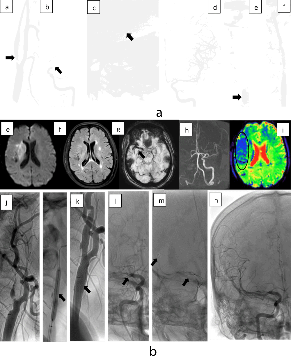
1a Distal approach. 62 years old patient with acute left T.O. (a) Diagnostic DSA with severe stenosis in the bulbar ICA with fl oating thrombus, (b) intracranial DSA showed thrombus, (c) Trevo 6/20 mm in the left MCA and ICA, (d) After the First pass, TICI 3 scale was achieved, (e) Balloon angioplasty was done in the ICA, (f) stent angioplasty was done. 1b. Proximal approach. 48 years old patient with acute right T.O. Diagnostic MRI (e) right the DWI showed a basal ganglia and subinsular white matter substance restriction area with (f) mismatch DWI/FLAIR, (g) SWI with thrombus in right M1, (h) The TOF MRA showed lack of signal in the right ICA and MCA s, (i) PWI with Tmax prolongation. (j) Pre dilatation balloon angioplasty was done with 3/15 mm and (k) a 7 mm x 40 cm carotid wall was used, (l) in the intracranial DSA proximal M1 was occluded, (m) a solitaire 4/20 mm was used, after two passes, (n) TICI 3 was achieved.
Read Full Article HTML DOI: 10.29328/journal.jnnd.1001051 Cite this Article Read Full Article PDF
More Images
Similar Articles
-
Analysis of early Versus Delayed Carotid Surgery after Acute Ischemic StrokePEROU Sébastien*,DETANTE Olivier,SPEAR Rafaelle,PIRVU Augustin,ELIE Amandine,MAGNE Jean-Luc. Analysis of early Versus Delayed Carotid Surgery after Acute Ischemic Stroke. . 2017 doi: 10.29328/journal.jnnd.1001001; 1: 001-011
-
Endovascular treatment experience in acute ischemic strokeİbrahim Acır*,Vildan Yayla,Hacı Ali Erdoğan. Endovascular treatment experience in acute ischemic stroke. . 2021 doi: 10.29328/journal.jnnd.1001047; 5: 026-028
-
Endovascular management of tandem occlusions in stroke: Treatment strategies in a real-world scenarioJuan J Cirio*,Celina Ciardi,Matias Lopez,Esteban V Scrivano,Javier Lundquist,Ivan Lylyk,Nicolas Perez,Pedro Lylyk. Endovascular management of tandem occlusions in stroke: Treatment strategies in a real-world scenario. . 2021 doi: 10.29328/journal.jnnd.1001051; 5: 055-060
Recently Viewed
-
Success, Survival and Prognostic Factors in Implant Prosthesis: Experimental StudyEpifania Ettore*, Pietrantonio Maria, Christian Nunziata, Ausiello Pietro. Success, Survival and Prognostic Factors in Implant Prosthesis: Experimental Study. J Oral Health Craniofac Sci. 2023: doi: 10.29328/journal.johcs.1001045; 8: 024-028
-
Agriculture High-Quality Development and NutritionZhongsheng Guo*. Agriculture High-Quality Development and Nutrition. Arch Food Nutr Sci. 2024: doi: 10.29328/journal.afns.1001060; 8: 038-040
-
A Low-cost High-throughput Targeted Sequencing for the Accurate Detection of Respiratory Tract PathogenChangyan Ju, Chengbosen Zhou, Zhezhi Deng, Jingwei Gao, Weizhao Jiang, Hanbing Zeng, Haiwei Huang, Yongxiang Duan, David X Deng*. A Low-cost High-throughput Targeted Sequencing for the Accurate Detection of Respiratory Tract Pathogen. Int J Clin Virol. 2024: doi: 10.29328/journal.ijcv.1001056; 8: 001-007
-
A Comparative Study of Metoprolol and Amlodipine on Mortality, Disability and Complication in Acute StrokeJayantee Kalita*,Dhiraj Kumar,Nagendra B Gutti,Sandeep K Gupta,Anadi Mishra,Vivek Singh. A Comparative Study of Metoprolol and Amlodipine on Mortality, Disability and Complication in Acute Stroke. J Neurosci Neurol Disord. 2025: doi: 10.29328/journal.jnnd.1001108; 9: 039-045
-
Development of qualitative GC MS method for simultaneous identification of PM-CCM a modified illicit drugs preparation and its modern-day application in drug-facilitated crimesBhagat Singh*,Satish R Nailkar,Chetansen A Bhadkambekar,Suneel Prajapati,Sukhminder Kaur. Development of qualitative GC MS method for simultaneous identification of PM-CCM a modified illicit drugs preparation and its modern-day application in drug-facilitated crimes. J Forensic Sci Res. 2023: doi: 10.29328/journal.jfsr.1001043; 7: 004-010
Most Viewed
-
Evaluation of Biostimulants Based on Recovered Protein Hydrolysates from Animal By-products as Plant Growth EnhancersH Pérez-Aguilar*, M Lacruz-Asaro, F Arán-Ais. Evaluation of Biostimulants Based on Recovered Protein Hydrolysates from Animal By-products as Plant Growth Enhancers. J Plant Sci Phytopathol. 2023 doi: 10.29328/journal.jpsp.1001104; 7: 042-047
-
Sinonasal Myxoma Extending into the Orbit in a 4-Year Old: A Case PresentationJulian A Purrinos*, Ramzi Younis. Sinonasal Myxoma Extending into the Orbit in a 4-Year Old: A Case Presentation. Arch Case Rep. 2024 doi: 10.29328/journal.acr.1001099; 8: 075-077
-
Feasibility study of magnetic sensing for detecting single-neuron action potentialsDenis Tonini,Kai Wu,Renata Saha,Jian-Ping Wang*. Feasibility study of magnetic sensing for detecting single-neuron action potentials. Ann Biomed Sci Eng. 2022 doi: 10.29328/journal.abse.1001018; 6: 019-029
-
Pediatric Dysgerminoma: Unveiling a Rare Ovarian TumorFaten Limaiem*, Khalil Saffar, Ahmed Halouani. Pediatric Dysgerminoma: Unveiling a Rare Ovarian Tumor. Arch Case Rep. 2024 doi: 10.29328/journal.acr.1001087; 8: 010-013
-
Physical activity can change the physiological and psychological circumstances during COVID-19 pandemic: A narrative reviewKhashayar Maroufi*. Physical activity can change the physiological and psychological circumstances during COVID-19 pandemic: A narrative review. J Sports Med Ther. 2021 doi: 10.29328/journal.jsmt.1001051; 6: 001-007

HSPI: We're glad you're here. Please click "create a new Query" if you are a new visitor to our website and need further information from us.
If you are already a member of our network and need to keep track of any developments regarding a question you have already submitted, click "take me to my Query."


