Figure 1
Post-stroke dizziness of visual-vestibular cortices origin
Nobuhiro Inoue* and Satoshi Goto
Published: 27 November, 2020 | Volume 4 - Issue 2 | Pages: 075-078

Figure 1:
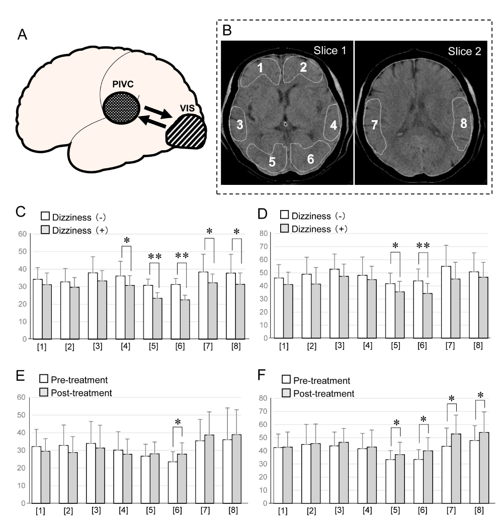
A: Schematic caricature demonstrated the relationship between PIVC and VISC concerned with dizziness in the patients with stroke. PIVC (Parieto Insular Vestibular Cortex), VISC (Visual Cortex).
B: Sections analyzed on CT images. Slice 1 and 2 are axial imaged that include the basal ganglia and temporo-parietal cortex respectively. ROIs were selected on each image. Slice 1: The level of the analyzed slice passed through the basal ganglia and included the midsectin of the anterior horns of the lateral ventricles, the caudate putamen, thalamus, and pineal body posteriorly. Three pairs of cortices corresponding to the frontal cortex ([1] and [2]), the temporal cortex ([3] and [4]), and the occipital cortex ([5] and [6]) = VISC were assessed. Slice 2: [7] and [8] represent the temporo-parietal cortex including the parieto-insular vestibular cortex (PIVC).
C: Comparison of the cerebral blood flow between the patients with dizziness and without dizziness at rest. Blank bar: cerebral blood flow in the Patients without dizziness. Gray bar: cerebral blood flow in the patients with dizziness. Significant difference between without dizziness and with Dizziness (*p < 0.05, **p < 0.01) appeared in [4],[5],[6], and [7],[8].
D: Comparison of the cerebral blood flow between the patients with dizziness and without dizziness after acetazolamide loading. Blank bar: cerebral blood flow in the patients without dizziness. Gray bar: cerebral blood flow in the patients with dizziness. Significant difference between without dizziness and with dizziness (*p < 0.05, **p < 0.01) appeared in [5] and [6].
E: Comparison of the cerebral blood flow between the patients with pre-treatment and post-treatment of Ibudilast at rest. Blank bar: cerebral blood flow in the patients with pre-treatment of Ibudilast. Gray bar: cerebral blood flow in the patients with post-treatment of Ibudilast. Significant difference between without dizziness and with dizziness (*p < 0.05) appeared only in [6].
F: Comparison of the cerebral blood flow between the patients with pre-treatment and post-treatment of Ibudilast after acetazolamide loading. Blank bar: cerebral blood flow in the patients with pre-treatment of Ibudilast. Gray bar: cerebral blood flow in the patients with post-treatment of Ibudilast. Significant difference between without dizziness and with dizziness (*p < 0.05) appeared in [5],[6],[7],and [8].
Read Full Article HTML DOI: 10.29328/journal.jnnd.1001038 Cite this Article Read Full Article PDF
More Images
Similar Articles
-
Analysis of early Versus Delayed Carotid Surgery after Acute Ischemic StrokePEROU Sébastien*,DETANTE Olivier,SPEAR Rafaelle,PIRVU Augustin,ELIE Amandine,MAGNE Jean-Luc. Analysis of early Versus Delayed Carotid Surgery after Acute Ischemic Stroke. . 2017 doi: 10.29328/journal.jnnd.1001001; 1: 001-011
-
Protective functions of AEURA in Cell Based Model of Stroke and Alzheimer diseaseJigar Modi,Ahmed Altamimi,Ashleigh Morrell,Hongyuan Chou,Janet Menzie,Andrew Weiss,Michael L Marshall, Andrew Li,Howard Prentice*,Jang-Yen Wu*. Protective functions of AEURA in Cell Based Model of Stroke and Alzheimer disease. . 2017 doi: 10.29328/journal.jnnd.1001003; 1: 016-023
-
Lateralized Cerebral Amyloid Angiopathy presenting with recurrent Lacunar Ischemic StrokeYi Li*, Ayman Al-Salaimeh,Elizabeth DeGrush,Majaz Moonis*. Lateralized Cerebral Amyloid Angiopathy presenting with recurrent Lacunar Ischemic Stroke. . 2017 doi: 10.29328/journal.jnnd.1001005; 1: 029-032
-
Comparative study of carboxylate and amide forms of HLDF-6 peptide: Neuroprotective and nootropic effects in animal models of ischemic strokeАnna P Bogachuk*,Zinaida I Storozheva,Andrey Т Proshin,Vyacheslav V Sherstnev,Irina V Smirnova,Tatyana M Shuvaeva,Valery M Lipkin. Comparative study of carboxylate and amide forms of HLDF-6 peptide: Neuroprotective and nootropic effects in animal models of ischemic stroke. . 2019 doi: DOI: 10.29328/journal.jnnd.1001022; 3: 096-101
-
Post-stroke dizziness of visual-vestibular cortices originNobuhiro Inoue*,Satoshi Goto. Post-stroke dizziness of visual-vestibular cortices origin. . 2020 doi: 10.29328/journal.jnnd.1001038; 4: 075-078
-
High suspicion of paradoxical embolism due to atrial septal Defect: A rare cause of ischemic strokeZahide Betul Gunduz*,Turgut Uygun. High suspicion of paradoxical embolism due to atrial septal Defect: A rare cause of ischemic stroke. . 2020 doi: 10.29328/journal.jnnd.1001040; 4: 084-087
-
Impact of mandibular advancement device in quantitative electroencephalogram and sleep quality in mild to severe obstructive sleep apneaCuspineda-Bravo ER*,García- Menéndez M,Castro-Batista F,Barquín-García SM,Cadelo-Casado D,Rodríguez AJ,Sharkey KM. Impact of mandibular advancement device in quantitative electroencephalogram and sleep quality in mild to severe obstructive sleep apnea. . 2020 doi: 10.29328/journal.jnnd.1001041; 4: 088-098
-
Cortical spreading depolarizations in the context of subarachnoid hemorrhage and the role of ketamineLeandro Custódio do Amaral*. Cortical spreading depolarizations in the context of subarachnoid hemorrhage and the role of ketamine. . 2021 doi: 10.29328/journal.jnnd.1001045; 5: 016-021
-
Endovascular treatment experience in acute ischemic strokeİbrahim Acır*,Vildan Yayla,Hacı Ali Erdoğan. Endovascular treatment experience in acute ischemic stroke. . 2021 doi: 10.29328/journal.jnnd.1001047; 5: 026-028
-
Factors associated with mortality after decompressive craniectomy in large basal ganglia bleedsAmit Kumar Thotakura*,Nageswara Rao Marabathina,Rama Krishnareddy Mareddy,Sivaramanjaneyulu Yeddanapudi . Factors associated with mortality after decompressive craniectomy in large basal ganglia bleeds. . 2021 doi: 10.29328/journal.jnnd.1001048; 5: 029-033
Recently Viewed
-
The Accuracy of pHH3 in Meningioma Grading: A Single Institution StudyMansouri Nada1, Yaiche Rahma*, Takout Khouloud, Gargouri Faten, Tlili Karima, Rachdi Mohamed Amine, Ammar Hichem, Yedeas Dahmani, Radhouane Khaled, Chkili Ridha, Msakni Issam, Laabidi Besma. The Accuracy of pHH3 in Meningioma Grading: A Single Institution Study. Arch Pathol Clin Res. 2024: doi: 10.29328/journal.apcr.1001041; 8: 006-011
-
Assessment of Perceptions of Nursing Undergraduates towards Mental Health PracticesAlya Algamdii*. Assessment of Perceptions of Nursing Undergraduates towards Mental Health Practices. Clin J Nurs Care Pract. 2025: doi: 10.29328/journal.cjncp.1001059; 9: 007-011
-
Multipurpose Antioxidants based on Food Industry Waste: Production and Properties EvaluationToshkhodjaev*. Multipurpose Antioxidants based on Food Industry Waste: Production and Properties Evaluation. Arch Food Nutr Sci. 2025: doi: 10.29328/journal.afns.1001062; 9: 001-003
-
Relationship between Fertility Diet Score Index Items and Ovulation in Women with Polycystic Ovary Syndrome: A Narrative ReviewHadis Alimoradi,Faezeh Mashhadi,Ava Hemmat,Mohsen Nematy,Maryam Khosravi,Maryam Emadzadeh,Nayere Khadem Ghaebi,Fatemeh Roudi*. Relationship between Fertility Diet Score Index Items and Ovulation in Women with Polycystic Ovary Syndrome: A Narrative Review. Arch Food Nutr Sci. 2024: doi: 10.29328/journal.afns.1001061; 8: 041-048
-
Evaluation of the LumiraDx SARS-CoV-2 antigen assay for large-scale population testing in SenegalMoustapha Mbow*,Ibrahima Diallo,Mamadou Diouf,Marouba Cissé#,Moctar Gningue#,Aminata Mboup,Nafissatou Leye,Gora Lo,Yacine Amet Dia,Abdou Padane,Djibril Wade,Josephine Khady Badiane,Oumar Diop,Aminata Dia,Ambroise Ahouidi,Doudou George Massar Niang,Babacar Mbengue,Maguette Dème Sylla Niang,Papa Alassane Diaw,Tandakha Ndiaye Dieye,Badara Cisé,El Hadj Mamadou Mbaye,Alioune Dieye,Souleymane Mboup. Evaluation of the LumiraDx SARS-CoV-2 antigen assay for large-scale population testing in Senegal. Int J Clin Virol. 2022: doi: 10.29328/journal.ijcv.1001041; 6: 001-006
Most Viewed
-
Feasibility study of magnetic sensing for detecting single-neuron action potentialsDenis Tonini,Kai Wu,Renata Saha,Jian-Ping Wang*. Feasibility study of magnetic sensing for detecting single-neuron action potentials. Ann Biomed Sci Eng. 2022 doi: 10.29328/journal.abse.1001018; 6: 019-029
-
Evaluation of In vitro and Ex vivo Models for Studying the Effectiveness of Vaginal Drug Systems in Controlling Microbe Infections: A Systematic ReviewMohammad Hossein Karami*, Majid Abdouss*, Mandana Karami. Evaluation of In vitro and Ex vivo Models for Studying the Effectiveness of Vaginal Drug Systems in Controlling Microbe Infections: A Systematic Review. Clin J Obstet Gynecol. 2023 doi: 10.29328/journal.cjog.1001151; 6: 201-215
-
Prospective Coronavirus Liver Effects: Available KnowledgeAvishek Mandal*. Prospective Coronavirus Liver Effects: Available Knowledge. Ann Clin Gastroenterol Hepatol. 2023 doi: 10.29328/journal.acgh.1001039; 7: 001-010
-
Causal Link between Human Blood Metabolites and Asthma: An Investigation Using Mendelian RandomizationYong-Qing Zhu, Xiao-Yan Meng, Jing-Hua Yang*. Causal Link between Human Blood Metabolites and Asthma: An Investigation Using Mendelian Randomization. Arch Asthma Allergy Immunol. 2023 doi: 10.29328/journal.aaai.1001032; 7: 012-022
-
An algorithm to safely manage oral food challenge in an office-based setting for children with multiple food allergiesNathalie Cottel,Aïcha Dieme,Véronique Orcel,Yannick Chantran,Mélisande Bourgoin-Heck,Jocelyne Just. An algorithm to safely manage oral food challenge in an office-based setting for children with multiple food allergies. Arch Asthma Allergy Immunol. 2021 doi: 10.29328/journal.aaai.1001027; 5: 030-037

HSPI: We're glad you're here. Please click "create a new Query" if you are a new visitor to our website and need further information from us.
If you are already a member of our network and need to keep track of any developments regarding a question you have already submitted, click "take me to my Query."


