Figure 1
Cranioplasty with preoperatively customized Polymethyl-methacrylate by using 3-Dimensional Printed Polyethylene Terephthalate Glycol Mold
Mehmet Beşir Sürme*, Omer Batu Hergunsel, Bekir Akgun and Metin Kaplan
Published: 30 November, 2018 | Volume 2 - Issue 2 | Pages: 052-064

Figure 1:
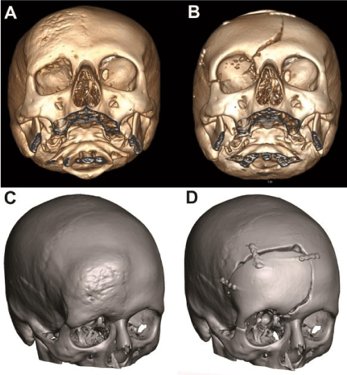
Preoperative (A) 3D CT and (C) Meshmixer software images of calvarium showing sclerotic thickening of right frontoorbital calvarial bone with narrowing of the orbital cavity. Postoperative (B) 3D CT and (D) Meshmixer software images of calvarium with patient-tailored PMMA implant. Following the resection of the lesion, frontal convexity, orbital roof and a part of the zygomatic bone is reconstructed with the prefabricated bone graft.
Read Full Article HTML DOI: 10.29328/journal.jnnd.1001016 Cite this Article Read Full Article PDF
More Images
Similar Articles
-
Cranioplasty with preoperatively customized Polymethyl-methacrylate by using 3-Dimensional Printed Polyethylene Terephthalate Glycol MoldMehmet Beşir Sürme*,Omer Batu Hergunsel,Bekir Akgun,Metin Kaplan. Cranioplasty with preoperatively customized Polymethyl-methacrylate by using 3-Dimensional Printed Polyethylene Terephthalate Glycol Mold. . 2018 doi: 10.29328/journal.jnnd.1001016; 2: 052-064
Recently Viewed
-
The Role of Anemia of Inflammation in the Course of Chronic HBV Infection in ChildrenInoyatova Flora Il’yasovna,Ikramova Nodira Anvarovna,Inogamova Gul’noza Zakhidjanovna,Kadyrkhodjayeva Khilola Marufovna,Abdullayeva Feruza Gafurovna,Valiyeva Nargiza Kabildjanovna. The Role of Anemia of Inflammation in the Course of Chronic HBV Infection in Children. J Adv Pediatr Child Health. 2025: doi: ; 8: 018-022
-
Forensic Perspectives on Human Chimerism: Identification Challenges and Detection StrategiesHarsh Kumar,Saumya Tripathi*. Forensic Perspectives on Human Chimerism: Identification Challenges and Detection Strategies. J Forensic Sci Res. 2025: doi: 10.29328/journal.jfsr.1001095; 9: 150-154
-
Minds after Death: The Expanding Role of Psychological Autopsy in Investigations: A ReviewIshan Jain*,Oindrila Mahapatra,Yogesh Kumar. Minds after Death: The Expanding Role of Psychological Autopsy in Investigations: A Review. J Forensic Sci Res. 2025: doi: 10.29328/journal.jfsr.1001096; 9: 155-0
-
Necrotizing Fasciitis in Neonates Case SeriesSaugat Ghosh*. Necrotizing Fasciitis in Neonates Case Series. J Adv Pediatr Child Health. 2025: doi: ; 8: 015-017
-
Effect of Azithromycin on Lung Function and Pulmonary Exacerbations in Children with Post-infectious Bronchiolitis Obliterans. A Double-blind, Placebo-controlled TrialCastaños Claudio*, Salin Maximiliano Felix, Pereyra Carla Luciana, Aguerre Veronica, Lucero Maria Belen, Bauer Gabriela, Zylbersztajn Brenda, Leviled Leonor, Gonzalez Pena Hebe. Effect of Azithromycin on Lung Function and Pulmonary Exacerbations in Children with Post-infectious Bronchiolitis Obliterans. A Double-blind, Placebo-controlled Trial. J Pulmonol Respir Res. 2024: doi: 10.29328/journal.jprr.1001052; 8: 009-014
Most Viewed
-
Feasibility study of magnetic sensing for detecting single-neuron action potentialsDenis Tonini,Kai Wu,Renata Saha,Jian-Ping Wang*. Feasibility study of magnetic sensing for detecting single-neuron action potentials. Ann Biomed Sci Eng. 2022 doi: 10.29328/journal.abse.1001018; 6: 019-029
-
Evaluation of In vitro and Ex vivo Models for Studying the Effectiveness of Vaginal Drug Systems in Controlling Microbe Infections: A Systematic ReviewMohammad Hossein Karami*, Majid Abdouss*, Mandana Karami. Evaluation of In vitro and Ex vivo Models for Studying the Effectiveness of Vaginal Drug Systems in Controlling Microbe Infections: A Systematic Review. Clin J Obstet Gynecol. 2023 doi: 10.29328/journal.cjog.1001151; 6: 201-215
-
Prospective Coronavirus Liver Effects: Available KnowledgeAvishek Mandal*. Prospective Coronavirus Liver Effects: Available Knowledge. Ann Clin Gastroenterol Hepatol. 2023 doi: 10.29328/journal.acgh.1001039; 7: 001-010
-
Causal Link between Human Blood Metabolites and Asthma: An Investigation Using Mendelian RandomizationYong-Qing Zhu, Xiao-Yan Meng, Jing-Hua Yang*. Causal Link between Human Blood Metabolites and Asthma: An Investigation Using Mendelian Randomization. Arch Asthma Allergy Immunol. 2023 doi: 10.29328/journal.aaai.1001032; 7: 012-022
-
An algorithm to safely manage oral food challenge in an office-based setting for children with multiple food allergiesNathalie Cottel,Aïcha Dieme,Véronique Orcel,Yannick Chantran,Mélisande Bourgoin-Heck,Jocelyne Just. An algorithm to safely manage oral food challenge in an office-based setting for children with multiple food allergies. Arch Asthma Allergy Immunol. 2021 doi: 10.29328/journal.aaai.1001027; 5: 030-037

HSPI: We're glad you're here. Please click "create a new Query" if you are a new visitor to our website and need further information from us.
If you are already a member of our network and need to keep track of any developments regarding a question you have already submitted, click "take me to my Query."


