Figure 1
Lateralized Cerebral Amyloid Angiopathy presenting with recurrent Lacunar Ischemic Stroke
Yi Li*, Ayman Al-Salaimeh, Elizabeth DeGrush and Majaz Moonis*
Published: 30 August, 2017 | Volume 1 - Issue 1 | Pages: 029-032

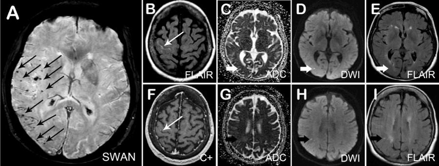
Figure 1:
Magnetic Resonance Imaging of lateralized cerebral amyloid angiopathy presented as recurrent ischemic stroke.
Susceptibility weighted angiography (SWAN) revealed diffuse scattered microhemorrhage lesions predominantly over the right MCA and PCA territory (A). FLAIR sulcal hyperintensity in the right central fi ssure with minimal enhancement was observed (B,F), indicating recent focal convexity subarachnoid hemorrhage or superfi cial cortical siderosis. Meanwhile, one DWI hyperintensive lesion over the right parietal lobe (D) was correlated with hypointensity signal on ADC (C) and isotensity signal on FLAIR (E); while another right parietal DWI hyperintensive
lesion (H) was correlated with almost isotensive signal on DWI (G) as well as hyperintensity on FLAIR (I), indicating lacunar infarctions at different ages.
Read Full Article HTML DOI: 10.29328/journal.jnnd.1001005 Cite this Article Read Full Article PDF
More Images
Similar Articles
-
Analysis of early Versus Delayed Carotid Surgery after Acute Ischemic StrokePEROU Sébastien*,DETANTE Olivier,SPEAR Rafaelle,PIRVU Augustin,ELIE Amandine,MAGNE Jean-Luc. Analysis of early Versus Delayed Carotid Surgery after Acute Ischemic Stroke. . 2017 doi: 10.29328/journal.jnnd.1001001; 1: 001-011
-
Lateralized Cerebral Amyloid Angiopathy presenting with recurrent Lacunar Ischemic StrokeYi Li*, Ayman Al-Salaimeh,Elizabeth DeGrush,Majaz Moonis*. Lateralized Cerebral Amyloid Angiopathy presenting with recurrent Lacunar Ischemic Stroke. . 2017 doi: 10.29328/journal.jnnd.1001005; 1: 029-032
-
Comparative study of carboxylate and amide forms of HLDF-6 peptide: Neuroprotective and nootropic effects in animal models of ischemic strokeАnna P Bogachuk*,Zinaida I Storozheva,Andrey Т Proshin,Vyacheslav V Sherstnev,Irina V Smirnova,Tatyana M Shuvaeva,Valery M Lipkin. Comparative study of carboxylate and amide forms of HLDF-6 peptide: Neuroprotective and nootropic effects in animal models of ischemic stroke. . 2019 doi: DOI: 10.29328/journal.jnnd.1001022; 3: 096-101
-
High suspicion of paradoxical embolism due to atrial septal Defect: A rare cause of ischemic strokeZahide Betul Gunduz*,Turgut Uygun. High suspicion of paradoxical embolism due to atrial septal Defect: A rare cause of ischemic stroke. . 2020 doi: 10.29328/journal.jnnd.1001040; 4: 084-087
-
Endovascular treatment experience in acute ischemic strokeİbrahim Acır*,Vildan Yayla,Hacı Ali Erdoğan. Endovascular treatment experience in acute ischemic stroke. . 2021 doi: 10.29328/journal.jnnd.1001047; 5: 026-028
-
Endovascular management of tandem occlusions in stroke: Treatment strategies in a real-world scenarioJuan J Cirio*,Celina Ciardi,Matias Lopez,Esteban V Scrivano,Javier Lundquist,Ivan Lylyk,Nicolas Perez,Pedro Lylyk. Endovascular management of tandem occlusions in stroke: Treatment strategies in a real-world scenario. . 2021 doi: 10.29328/journal.jnnd.1001051; 5: 055-060
-
Differential roles of trithorax protein MLL-1 in regulating neuronal Ion channelsSonya Dave,An Zhou*. Differential roles of trithorax protein MLL-1 in regulating neuronal Ion channels. . 2021 doi: 10.29328/journal.jnnd.1001057; 5: 089-093
-
Stroke Mimics: Insights from a Retrospective Neuroimaging StudyLucia Monti*, Davide del Roscio, Francesca Tutino, Tommaso Casseri, Umberto Arrigucci, Matteo Bellini, Maurizio Acampa, Sabina Bartalini, Carla Battisti, Giovanni Bova, Alessandro Rossi. Stroke Mimics: Insights from a Retrospective Neuroimaging Study. . 2023 doi: 10.29328/journal.jnnd.1001083; 7: 094-103
Recently Viewed
-
Development and Evaluation of a mHealth app - (ReMiT-MS app) for Rehabilitation of Individuals with Relapsing-remitting Multiple Sclerosis - A Mixed Methods, Pragmatic Randomized Controlled Trial - Study ProtocolSolaiyan Rajanchellappa, Dheeraj Khurana*, AGK Sinha, Soundappan Kathirvel, Ashok Kumar, Rajni Sharma. Development and Evaluation of a mHealth app - (ReMiT-MS app) for Rehabilitation of Individuals with Relapsing-remitting Multiple Sclerosis - A Mixed Methods, Pragmatic Randomized Controlled Trial - Study Protocol. J Nov Physiother Rehabil. 2024: doi: 10.29328/journal.jnpr.1001060; 8: 022-030
-
Enhancing Physiotherapy Outcomes with Photobiomodulation: A Comprehensive ReviewNivaldo Antonio Parizotto*, Cleber Ferraresi. Enhancing Physiotherapy Outcomes with Photobiomodulation: A Comprehensive Review. J Nov Physiother Rehabil. 2024: doi: 10.29328/journal.jnpr.1001061; 8: 031-038
-
From Adversity to Agency: Storytelling as a Tool for Building Children’s ResilienceKate Markland*. From Adversity to Agency: Storytelling as a Tool for Building Children’s Resilience. J Nov Physiother Rehabil. 2024: doi: 10.29328/journal.jnpr.1001062; 8: 039-042
-
Physiotherapy Undergraduate Students’ Perception About Clinical Education; A Qualitative StudyPravakar Timalsina*,Bimika Khadgi. Physiotherapy Undergraduate Students’ Perception About Clinical Education; A Qualitative Study. J Nov Physiother Rehabil. 2024: doi: 10.29328/journal.jnpr.1001063; 8: 043-052
-
Management of Non-contact Injuries, Nonspecific Chronic Pain, and Prevention via Sensory Conflicts Detection: Vertical Heterophoria as a Landmark IndicatorEric Matheron*. Management of Non-contact Injuries, Nonspecific Chronic Pain, and Prevention via Sensory Conflicts Detection: Vertical Heterophoria as a Landmark Indicator. J Nov Physiother Rehabil. 2024: doi: 10.29328/journal.jnpr.1001057; 8: 005-013
Most Viewed
-
Evaluation of Biostimulants Based on Recovered Protein Hydrolysates from Animal By-products as Plant Growth EnhancersH Pérez-Aguilar*, M Lacruz-Asaro, F Arán-Ais. Evaluation of Biostimulants Based on Recovered Protein Hydrolysates from Animal By-products as Plant Growth Enhancers. J Plant Sci Phytopathol. 2023 doi: 10.29328/journal.jpsp.1001104; 7: 042-047
-
Sinonasal Myxoma Extending into the Orbit in a 4-Year Old: A Case PresentationJulian A Purrinos*, Ramzi Younis. Sinonasal Myxoma Extending into the Orbit in a 4-Year Old: A Case Presentation. Arch Case Rep. 2024 doi: 10.29328/journal.acr.1001099; 8: 075-077
-
Feasibility study of magnetic sensing for detecting single-neuron action potentialsDenis Tonini,Kai Wu,Renata Saha,Jian-Ping Wang*. Feasibility study of magnetic sensing for detecting single-neuron action potentials. Ann Biomed Sci Eng. 2022 doi: 10.29328/journal.abse.1001018; 6: 019-029
-
Pediatric Dysgerminoma: Unveiling a Rare Ovarian TumorFaten Limaiem*, Khalil Saffar, Ahmed Halouani. Pediatric Dysgerminoma: Unveiling a Rare Ovarian Tumor. Arch Case Rep. 2024 doi: 10.29328/journal.acr.1001087; 8: 010-013
-
Physical activity can change the physiological and psychological circumstances during COVID-19 pandemic: A narrative reviewKhashayar Maroufi*. Physical activity can change the physiological and psychological circumstances during COVID-19 pandemic: A narrative review. J Sports Med Ther. 2021 doi: 10.29328/journal.jsmt.1001051; 6: 001-007

HSPI: We're glad you're here. Please click "create a new Query" if you are a new visitor to our website and need further information from us.
If you are already a member of our network and need to keep track of any developments regarding a question you have already submitted, click "take me to my Query."


